Brynna presented to our Soft Tissue Surgery Service with a history of a draining sinus located over her left flank.
She had undergone a previous surgery prior to referral to treat a suspect abscess located on the left thigh. Culture of the tissue was negative and no neoplastic process was identified on histopathology. Initially we were suspicious of a migrating foreign body as the primary cause of these lesions.
We decided to perform a positive contrast CT scan of the thorax, abdomen and pelvic limb. We also performed a sinogram: injection of contrast into the sinus to outline and define any communications of the draining sinus and a potential foreign body.
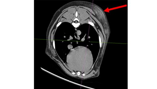
This CT scan identified three lesions – the draining wound on her flank (which communicated to a suspected abscess under her spine), a nodule in her armpit/axilla and a third mass immediately behind her scapula.
We elected to proceed with surgical exploration; exploratory laparotomy, debridement of the left flank lesion, and excision biopsy of the other two lesions. No foreign body was identified and tissue was submitted for both histopathology and culture.
No bacterial or fungal growth was noted after 72 hour culture. Histopathology was consistent with sterile nodular panniculitis.
Three days after surgery Brynna was noted to have some pain/discomfort eating food and was not interested in playing with her ball.
Although not confirmed, we suspect that this oral pain was related to an inflammatory myopathy as part of this immune mediated process.
Based on the histopathology findings Brynna was started on an immunosuppressive course of prednisolone (2mg/kg/day). She has responded very well to this therapy and her oral discomfort has resolved.
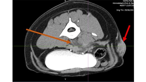
Left image: Orange area – fluid pocket ventral to spinal vertebrae, Red area – subcutaneous mass on left flank
Right image: Mass behind left shoulder